Photo A
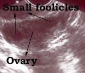
A normal ovary. A few small follicles are visible (it is completely normal)
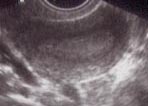
Photo B

A normal ovary with a follicle. A egg cell ready for release will be present in the follicle. This is not a cyst.
Photo B
A normal ovary with a follicle. A egg cell ready for release will be present in the follicle. This is not a cyst.
Photo C

This is a ovary with multiple irregular cysts and turned out to a cystic cancer of the ovary. This photo is included to stress the importance of regular checks. Regular annual examinations and regular ultrasound examinations done through the vagina might help to diagnose ovarian cancer early.
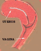
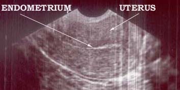
The transvaginal sonar also give an excellent view of the cervix ( mouth of the womb) helping doctors to excess conditions effecting the cervix.
We tried to explain the increasing importance of a transvaginal ultrasound (sonar) examinations in assisting doctors to access female (gynecological) diseases.
We will now discuss the use of ultrasound in pregnancy.
The first concern is the safety to the fetus. Until now no dangerous side effects or abnormalities caused by sonar examination has been discovered.
The role of sonar examinations during pregnancy is to detect the duration of pregnancy (dating the pregnancy), to detect fetal abnormalities and to monitor fetal development and growth.
It is important to understand that the sonar is just a machine and can't determine the duration of pregnancy. What actually happens is that different fetal parts are measured and that according the size of the specific part a computer program calculates the duration of the pregnancy. The programs are developed by doing thousands of ultrasound examinations at different stages of pregnancy. The accuracy of sonar dating of a pregnancy depends on different factors.
Is the program appropriate to the community where it is used. If the program was developed in a country where the average fetal size is small and used in community where the average fetal size is large ( or vice versa) unreliable results will be obtained. A fetus in the population with the large average size will appear to bigger and older than what it actually is. The opposite will happen in the population with the smaller than average size fetus.
The size of a fetus also influences the accuracy of a ultrasound's estimation of the duration of pregnancy. A larger than average fetus will cause the program to abnormally increase the duration and a small fetus will cause the opposite effect.
Here are a few sonar photos to illustrate the appearance of the fetus at different stages of a pregnancy.
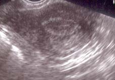
Photo One
This a six week pregnancy and a transvaginal probe was used.
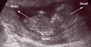
Photo Two
This is a nine weeks pregnancy as seen using a vaginal probe.
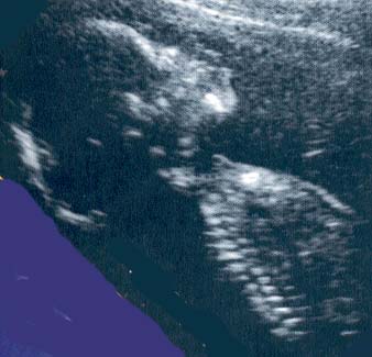
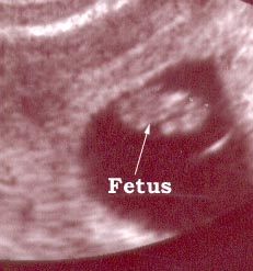
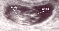
Photo Three
This is a twelve weeks pregnancy as seen by external (through the abdomen) sonar.
Another point to remember is that the reference point used on ultrasound machines to determine the duration of pregnancy is the first day of the last menstruation and NOT THE MOMENT OF CONCEPTION.
There are two reference points used to determine the duration of pregnancy :
(i) The first day of the last menstruation.
(ii) The moment of conception.
The fist is in use for centuries because the only known fact was that menstruation stopped during pregnancy
Than a new scientific division known as embryology developed. Embryology is the study of development before birth. Embryologists originally examined animal embryos with a known date of conception. Hence the moment of conception became their reference point. Human embryologists also use the moment of conception as their reference point, Obstetricians however still use the first day of the last menstruation as their reference point. This can lead to confusion because conception occurs about 2 (two) weeks after the first day of the last menstruation.
The reference point use also has legal implications in fraternity disputes. If a sonar examination indicates a twelve week pregnancy , the duration is only 10 weeks and conceptions occurred about 10 weeks ( and not twelve weeks) previously.
|